Study Tips for the Final Exam

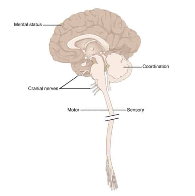
Anatomical Underpinnings of the Neurological Exam
Anatomical Underpinnings of the Neurological Exam (source Anatomy & Physiology OpenStax College; Download for free at http://cnx.org/content/col11496/latest/)
Study Tip for the Final Exam 1.

Prof. White gives very important Study Tips for the Final Exam in the video “Preparing for the Comprehensive Final Exam”. In addition to that  a special training exam has been prepared. You can find it in the Quizzes and Exams. It is called Final Exam Preparation Example Clinical Case Quiz. This is ungraded exam and allows you to practice for the Final Exam.

Study Tip for the Final Exam 2.

Again use past material to review.  If you go to the list of Video Tutorials on the course website, you find two files subtitles (txt) and subtitles (srt) with every tutorial. You can use the .txt files from the tutorials that contain information on the concept you want to review to decide which of the tutorials you want to watch again.

The subtitles (srt) refers also to the timeslot in the tutorial where the text is spoken. That allows you to go back to a specific time in a tutorial and review the visual material that is presented on a certain neuromedical concept.

Study Tip for the Final Exam 3.

The Comprehensive Final Exam has 4 parts:

  • Part 1 (2 cases, 13 questions).
  • Part 2 (2 cases, 7 questions).
  • Part 3 (2 cases, 11 questions).
  • Part 4 (2 cases, 12 questions).
Study Tip for the Final Exam 4.
  • 3-D Brain app by Cold Spring Harbor Laboratory. A very useful 3D model of the brain. It has descriptions of various brain structures, associated functions, associated cognitive disorders, etc. It is useful for the preparation of Quizzes and especially relevant for the Comprehensive Final Exam. It was accessible with the internet browser, but that site used  Flash, it does not work anymore. You have to download the App. You can get the free 3D Brain app from Google play and Windows Phone and the App Store from Apple.
Study Tip for the Final Exam 5.

On the Virtual Lab, Unit 5: Cognition you can find another useful resource: Regions and functions in the cerebral cortex, a list of functions and deficits or problems revealed when injury occurs at particular locations, by Centre for Neuro Skills.

Study Tip for the Final Exam 6.

Reminder, in Study Tips Sensory Systems this tip can be found:  There is a place in the brainstem where damage can lead to dissociated sensory loss in sensations to the face. In medical neuroscience it is important to be able attribute this dissociated sensory loss to the structures in the medulla. It could be very useful to re-watch tutorial Pain and Temperature Pathways (part 2) from 15.00 to the end of the tutorial.

Study Tip for the Final Exam 7.

On the Virtual lab this link can be found MRI images: MRI  – Frontal,  – Axial, – Saggital (move the cursor over the image to see the labels).

Study Tip for the Final Exam 8.

At the page Neurological Exam there are very useful resources in the paragraphs: Neurological Exam, NeuroLogical Cases and Clinical Neurology Videos. Use them well to prepare for the final exam.

Study Tip for the Final Exam 9.

Left and right in axial images. Because most of the images are quite symmetrical, as is the entire human body, it is obviously important to know where is left and right side of the actual patient.  Especially so for Clinical Cases and the exams, where you’ll need to relate presented symptoms to anomalities on an image.  Brainstem and spinal cord slices as demonstrated in Sylvius, and the tutorial notes are presented in axial view. The point of view is from below. It is like we are looking to the persons top of the head through his or her feet. It is the same as for Magnetic Resonance Images as for photographs and drawings.

There is no universal standard of orientation of the brain images, but the most common convention is as follows:

  • Photographic images or drawings: with the anterior side below and the posterior side above. It is like the person is lying on his/her belly, face downward. What you see on the left side is at the left side of the body.
  • MRI images: with the anterior side above and the posterior side below. It is like the person is lying on his/her back.  What you see on the left side is at the right side of the body and vice versa.
Study Tip for the Final Exam 10

When reviewing the material of the course the Summary of Pathways  (2020, Neuroscience 5th and 6th ed.) This is a summary for the Final Exam of the course. Images in the Summary come from Anatomy & Physiology (OpenStax College .© 1999-2016, Rice University) is very useful.. You can use this material for educational purposes but remember to give the source. You can look at Virtual Lab, General Resources to find out how to do that.
Study Tips Cognition

Study Tips Cognition

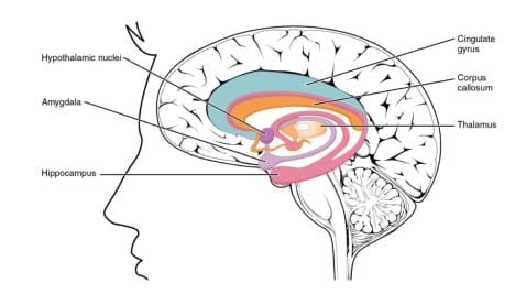
limbic structures
Limbic Structures (source Anatomy & Physiology OpenStax College; Download for free at http://cnx.org/content/col11496/latest/)

There is  a new, 6th edition of ‘Neuroscience’ , the course textbook. It can be used instead of the 5th edition that is often used.

The part in the course on Cognition will be enriched  in the future by another resource. Prof. White will add content from the second edition of Dr. Purves’ and colleagues’ other major textbook, Principles of Cognitive Neuroscience . 

Consequently the resources and the Study Tips on Cognition, will be updated to the new content.

Study Tip Cognition 1.

In Brain Development you had an excellent opportunity to review material. You have the same opportunity in Cognition.

First of all there is this strategic Study Tip for reviewing previous material.

Under a Video Tutorial at the left hand side of your screen you see in the list of Downloads two text files. The first one is Transcript (txt).

You can use the .txt files from the tutorials that contain information on the concept you want to review to decide which of the tutorials you want to watch again. You can open the txt files in Notebook.

The information in the file ‘Subtitles’ (WEBVTT)) refers also to the time slot in the tutorial where the text is spoken. You can go back to a specific time in a tutorial and review the visual material that is presented on a certain neuromedical concept.

Study Tip Cognition 2.

Also you can use the resources on the Virtual Lab, Unit 5: Cognition.

Study Tip Cognition 3.

Previously introduced resources are also very important in Cognition, like:

Anatomical aspects of cognition: 

Psychological aspects of cognition:

Cognition in relation to deficits, damage and disease:

Limbic Forebrain:


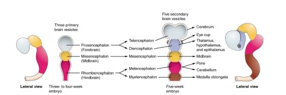
Study Tips Brain Development

brain development
Brain development (source Anatomy & Physiology OpenStax College; Download for free at http://cnx.org/content/col11496/latest/)

The Study Tips Brain Development is not a very rich page on the website compared to previous pages. Most of the strategic Study Tips are present on earlier pages and are not repeated here. Especially Brain anatomy is important in this Unit of the course. Relevant Study Tips and resources for Brain Development are presented in Study Tips Neuroanatomy and in the Virtual Lab, Unit A: Neuroanatomy.

Study Tip Brain Development 1.

This course starts with Neuroanatomy. Brain Development comes later in the course. Therefore you can review a lot of the material of the Neuroanatomy unit in the unit on Brain Development.

You can go back to the Unit A tutorial notes (even better re-watch Unit A tutorials if you have time to spare) and review Neuroanatomy for your study of Brain Development. For example, prof. White presents an embryological framework for cranial nerve nuclei in the tutorial notes on Cranial Nerve Nuclei. You can review this material and expand your understanding of embryological development at the same time.

Study Tip Brain Development 2.

I find Figure: The ventricular spaces associated with each of the major subdivisions of the brain (A24 page 744 of the 5th edition and A2 on page A-3 of the 6th edition of the textbook) very useful in the study of this Unit.

Study Tip Brain Development 3.

You can use the resources on the Virtual Lab, Unit 4: Brain Development.

Study Tip Brain Development 4.

Besides course material there is more relevant material in the next list of resources

Early Brain Development.
Modification of Neural Circuits in Early Neonatal Life
The Changing Brain Across the Lifespan; development, repair and regeneration

Study Tips for the Functional Anatomy Exam

Study Tips for the Functional Anatomy Exam

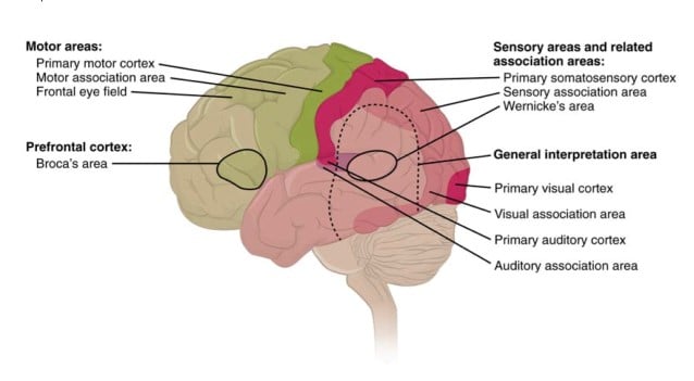
types of cortical areas
Types of Cortical Areas (source Anatomy & Physiology OpenStax College; Download for free at http://cnx.org/content/col11496/latest/)

The Functional Anatomy Exam is not currently included in the assessment plan for Medical Neuroscience. The assessment plan might include it again in the future. The Study Tips can be valuable to general insight in Medical Neuroscience whether there is a Functional Anatomy Exam or not. For that reason they are included in the website.

Study Tip Functional Anatomy Exam 1.

The exam consists of several parts:

  • 15 questions on Surface Anatomy of the Brain
  • 6 questions on the Internal Anatomy of the Brainstem
  • 4 questions on the Internal Anatomy of the Spinal Cord
  • 5 questions on Motor Pathways
  • 5 questions on on Sensory Pathways
  • 5  questions on  Internal Anatomy of the Forebrain
Study Tip Functional Anatomy Exam 2

You are free to use the Summary of Pathways. You can use it and share it. You have to respect the attributions.

Study Tip Functional Anatomy Exam 3, Surface Anatomy of the Brain:

First of all you can use the unlabeled images of the brain for review that are presented in the tutorial notes:

  • Fig 1. Lateral surface of the human Brain in the tutorial on the Lateral Surface of the Brain, and the tutorial Finding the Central Sulcus
  • Fig.1. The Medial surface of the human brain in the corresponding tutorial, and the tutorial Finding the Central Sulcus
  • Fig 1. Ventral surface of the brain in the corresponding tutorial.

Besides this course material the site 3-D Brain App is especially relevant for your study of the Surface Anatomy of the Brain. Most noteworthy is the 3D-Brain on this site.  The 3D-Brain gives the anatomical place of a structure, its associated functions, associated cognitive disorders etc. It was accessible with the internet browser, but that site used Flash, it does not work anymore. You have to download the App. You can get the free 3D Brain app from Google play and Windows Phone and the App Store from Apple.

The site Functional Cortical Anatomy (Lateral Surface) by Handwritten Tutorials is also very good additional material for this part of the Functional Anatomy Exam. The video provides clear visual insight.

Study Tip Functional Anatomy Exam 4, Brainstem:
  • First of all you can study  tutorial notes: Internal Anatomy of the Brainstem and Cranial Nerve Nuclei (Unit A) thoroughly because the figures are very useful.
  • Damage in a certain  place in the brainstem can lead to dissociated sensory loss of sensations to the face. You must be able attribute this dissociated sensory loss to the structures in the medulla.
  • The anatomy of the brainstem is important and because of that it is useful to re-watch tutorial 3-16 from 15.36 to the end of the tutorial.
  • You can also use the cards provided by B.C. Jackson to revise the anatomy of the parts of the brainstem and spinal cord. These cards give you the opportunity to draw the relevant structures of the pathways and other brainstem structures.
  • If you do not Sylvius you can make use of:
Study Tip Functional Anatomy Exam 5, Internal Anatomy of the Spinal Cord:

The site Spinal Cord/Brainstem Interactive Atlas by Bellarmine University presents relevant additional information too.

Study Tip Functional Anatomy Exam 6, Motor and Sensory Pathways:

In addition to the Summary of Pathways you can use the table  and produce your own versions of pathway images using these images, In that way you will make a summary of pathways that you can use in the parts on Motor and Sensory pathways.

Study Tip Functional Anatomy Exam 7, Internal Anatomy of the Forebrain: 
  • You can use tutorial notes; Internal Capsule and Deep Gray Matter; Basal ganglia; In tutorial 4-18 at 8.29 to 11.02 the basal ganglia in the brainstem model are pointed out. It is a good material to get a grip on the spatial orientation of the basal ganglia.
Study Tip Functional Anatomy Exam 8.

In the Tutorial Professor White’s Favorite Places in the Human Brain Professor White shows his favorite places in the brain. It is a tutorial rich in relevant information that could have been a bit overpowering at the start of the course. Therefore you will be surprised to find out that you really understand what is presented. Consequently it is really useful to re-watch at this stage in the course.

Study Tip Functional Anatomy Exam 9.

Left and right in axial images. While most of the images are quite symmetrical, as is the entire human body, it is obviously important to know where is left and right side of the actual patient.  Especially so for Clinical Cases and the exams, where you’ll need to relate presented symptoms to anomalies on an image.  For example: Brainstem and spinal cord slices as demonstrated in Sylvius and the tutorial notes are presented in axial view. The point of view is from below, it is like we are looking to the persons top of the head through his or her feet. It is the same as for Magnetic Resonance Images as for photographs and drawings.


Study Tips Motor Systems

Study Tips Motor Systems

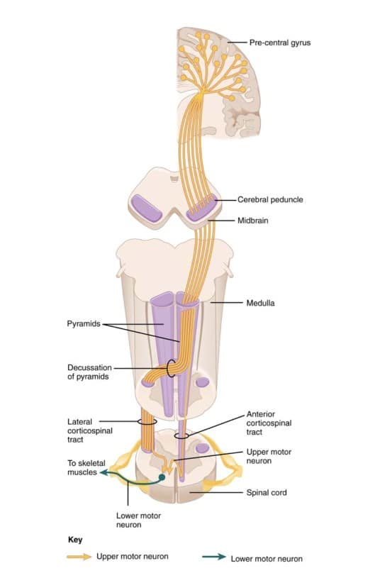
corticospinal tract
Corticospinal tract (source Anatomy & Physiology OpenStax College; Download for free at http://cnx.org/content/col11496/latest/)
Study Tip Motor Systems 1.

The sensory pathways have the purpose of getting the stimulus/action potential to the brain as fast and as efficient as possible. They are the highways of the nervous system. They share the 3 neuron ‘design.’

Motor pathways have to do with the execution of  behavior. They are more diverse than sensory pathways and are fine-tuned by other neural systems. Opposed to the sensory pathways finding comparable key elements to compare the anatomy is not feasible. In contrast with sensory pathways they do not have a 3 neuron ‘design’.

The most direct motor pathways are the ‘Direct Somatic Motor pathways’. The corticospinal tracts for execution of behavior in the body. And the corticobulbar tracts for execution of behavior in the  face. Other motor pathways are the indirect pathways, the brainstem–to-spinal cord pathways.

First of all you can have a look at the direct and indirect motor systems in this animation: The extrapyramidal and pyramidal motor systems .

Study Tip Motor Systems 2.

Also you can use  the resources on page Virtual Lab, Unit 3: Motor Systems to enrich your understanding of the core concepts of motor systems.

Study Tip Motor Systems 3.

Basal ganglia are very important in motor pathway. In the tutorial: ‘Circuitry of the Basal Ganglia, part 2  ‘ at 8.28 to 11.02 the basal ganglia in the brainstem model are pointed out. This video material of the brainstem is good material to get a grip on the spatial orientation of the basal ganglia. In addition to this valuable course material it is very good to use this site  of the basal ganglia, by the University of British Columbia. for your study. This animation: 2-Minute Neuroscience: Basal Ganglia by Neuroscientifically Challenged, further allows you to review the anatomy of the basal ganglia and ties it to the motor functions of the basal ganglia.

Study Tip Motor Systems 4.

In addition to your study of the course material you can study reflexes by having a good look at the relevant animations of the textbook:  Stretch reflex animation and  The flexion reflex.

Study Tip Motor Systems 5.

Finally the “Handwritten Tutorials” on YouTube are very useful in your study.  The ones in bold are especially relevant to this unit.

Study Tip Motor System 6.

Most relevant figures from the second edition of the course textbook for sensory and motor systems are below. You can copy the name of the figure and paste it in ‘Search in this book’. Then a list of relevant pages will appear. Click ‘Show details’ of the most relevant page and a link to the most relevant page appears, click that and scroll down to locate the figure. You can only use these images for your own study, images can’t be shared. Copyright:. D. Purves et al. 2001, Sinauer Associates, Inc. For more information, see the Bookshelf Copyright Notice.

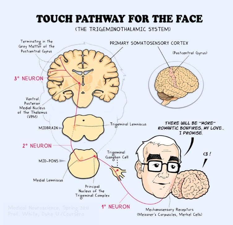
  • Dorsal column medial lemniscal pathway and Trigeminal lemniscal pathway= figure 9.6
  •  Anterolateral pathway/Spinothalamic tract and Trigeminal thalamic pathway = figure 10.3
  •  (Lateral and anterior) corticospinal tract. Pyramidal tracts = figure 17.9
  •  Retina-geniculate-striate cortex pathway = figure 12.8
  •  Pupillary light reflex = figure 12.3
  •  Direct and indirect pathway of the basal ganglia = figure 18.1
  •  Disinhibition in direct and indirect pathways of the basal ganglia in a healthy brain =figure 18.8
  •  Hypo and hyperkinetic disorders on movement control pathways = figure 18.10
  •  Input into cerebellum = figure 19.3
  •  Ascending output directed to thalamocortical circuits = figure 19.6
Study Tips Motor System 7.

It is a very good idea to  use the material on the page Learning Materials for reviewing pathways.

Study Tips Sensory Systems

Study Tips Sensory Systems

Brain Caress
Brain Caress. Image from the Peer Assessment of the Medical Neuroscience course in 2013
Study Tips Sensory Systems 1.

Now you are starting with the intense core of Medical Neuroscience, Units 3 and 4. First of all it is a good idea to start a thread in the forum to  form a study group and share your experiences. You can be in touch with fellow students. that  can help when the going gets tough. In addition to forum study groups you can also join the Medical Neuroscience Facebook group. That page was started in 2013 when Medical Neuroscience ran for the first time.It  is open to this class as well. That 1st year people were sharing their drawing efforts for the assessments. It was inspiring and great fun, there were real artist among us (see photos on Facebook page).

Study Tips Sensory Systems 2.

You can use the resources on the page Virtual Lab, Unit 2: Sensory Systems.

Study Tips Sensory Systems 3.

Drawing pathways is a powerful learning tool (see Week 5, Somatic sensation, Tutorial Mechanosensory pathways, part 3; 09.50-14.25 for Dr. White’s directions for learning pathways). You can draw pathways and share  your drawings in the learning community on the forum. It is very good to assess the attempts of your peers and give feedback on their images. As a result you both learn. 🙂

Study Tips Sensory Systems 4.

The tutorial notes contain a compact and strong overview  of pathways that is a very good resource for effective, active learning. The table can be found in the Tutorial Notes on Mechanosensory pathways,  page 8:

Iva Aleksova (former Teaching Assistant) has made the table available in Google Documents. You can follow the link and you can download it in Word so you can manipulate it to suit your requirements.  Here are some suggestions to use it for effective learning.

  • Customize the table.
  • You can supplement the table with images. Prints from Sylvius with printscreen (if you change preference, for example both for selected and unselected structures at 0.05 you can easily locate structures in the print). Or use the brainstem cards our mentor B.C. Jackson has made  to visualize parts of pathways.
  • You can create comparable tables when you make overviews of pain pathways.
Study Tips Sensory Systems 5.

For the visual system and auditory system this table cannot be used directly. Learners can try to adapt the table and write relevant information in a systematic, compact way. The product of this exercise may not be a beautiful table but instead an effective way to learn the key elements of these pathways. For central visual pathways it may be an idea to have 8 rows with quadrants of the visual fields (left superior temporal field, left superior nasal field, left inferior temporal field, left inferior nasal field, right……, etc). A row with information about the place in the optic radiation could be included for instance, maybe another about the place in the visual cortex.. The pupillary light reflex poses a challenge. Can you make  a table for that? What are key elements in that pathway? And can you make a table for the auditory and vestibular system?

Study Tips Sensory Systems 6.

In the course B.C. Jackson has created a resource with free images of structures important in pathways (called brainstem tract images). You are free to download those images and use them for your study .

Here are some suggestions:

  • You can draw the first, second and third order neuron for each pathway (What is the associated gray matter structure?)
  •  You can also draw the level of decussation in the pathway (Which neuron? Which structures are important?)
  •  Which part of the thalamus is part of the pathway?
  •  Where is the primary cortical area?
  •  Which important white matter components can be found in the pathway?
  •  Etc.
Study Tips Sensory Systems 7.

It is a very good idea to  use the material on the page Learning Materials for reviewing pathways.

Study Tips Sensory Systems 8.

There is a place in the brainstem where damage can lead to dissociated sensory loss in sensations to the face. In medical neuroscience it is important to be able to attribute this dissociated sensory loss to the structures in the medulla. It could be very useful to re-watch Week 5, Somatic sensation, Tutorial Pain and Temperature Pathways, part 2  from 15.30 to the end of the tutorial.

Study Tips Sensory Systems 9.

Use the cards provided by B.C. Jackson to revise the anatomy of the parts of the brainstem and spinal cord. Use them to draw the relevant structures of the pathways. Start drawing and reviewing other brainstem structures like cranial nuclei as well, which will prove very useful when reviewing for the exams.

Study Tips Sensory Systems 10.

Most relevant figures from the second edition of the course textbook for sensory and motor systems are below. You can copy the name of the figure and paste it in ‘Search in this book’.Then a list of relevant pages will appear. Click ‘Show details’ of the most relevant page and a link to the most relevant page appears, click that and scroll down to locate the figure. You can only use these images for your own study, images can’t be shared. Copyright:. D. Purves et al. 2001, Sinauer Associates, Inc. For more information, see the Bookshelf Copyright Notice.

  • Dorsal column medial lemniscal pathway and Trigeminal lemniscal pathway= figure 9.6
  •  Anterolateral pathway/Spinothalamic tract and Trigeminal thalamic pathway = figure 10.3
  •  (Lateral and anterior) corticospinal tract. Pyramidal tracts = figure 17.9
  •  Retina-geniculate-striate cortex pathway = figure 12.8
  •  Pupillary light reflex = figure 12.3
  •  Direct and indirect pathway of the basal ganglia = figure 18.1
  •  Disinhibition in direct and indirect pathways of the basal ganglia in a healthy brain =figure 18.8
  •  Hypo and hyperkinetic disorders on movement control pathways = figure 18.10
  •  Input into cerebellum = figure 19.3
  •  Ascending output directed to thalamocortical circuits = figure 19.6
Study Tips Sensory Systems 11.

I like the “Handwritten Tutorials” spinal series on YouTube.

Study Tips Neural Signaling

Study Tips Neural Signaling

action potential ( source anatomy & physiology)
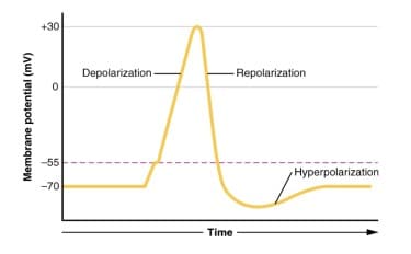
Action Potential (source Anatomy & Physiology OpenStax College; Download for free at http://cnx.org/content/col11496/latest/)

Concrete additions to your study strategy to make your study more successful and more fun with optimal use of the resources.

Study Tip Neural Signaling 1.

Use the resources in the Virtual Lab, Unit 1: Neural Signaling.

Study Tip Neural Signaling 2.

When you do not feel confident on your biological background, you can try if this book can be of help:  Biology – OpenStax college. Chapter 35: The Nervous System and Chapter 36: Sensory Systems

Study Tip Neural Signaling 3.

If you are watching a tutorial and think ‘hearing your explanation that makes sense’ but you are not sure you will recall the explanation when you get questions about it, write down the time and tutorial of the explanation in your notes. Then it’s easier to re-view the explanation to really get it.

Study Tip Neural Signaling 4.

For those of you that do not have the textbook, you can use the digital edition of the textbook (second edition). For example:

  1. An important figure is figure 5.3  Sequence of events involved in transmission at a typical chemical synapse. It is figure 5.3 in the second edition as well.
  2. An important figure is a figure on excitatory and inhibitory post synaptic potentials (figure 5.21 in the tutorial). It can be found in the second edition as well. There the number of the figure is 7.6.
  3. the figure on NMDA receptors (fig.8.10 in the tutorial) can be found as figure 25.9 in the digital edition of Neuroscience.

In this digital edition you can also read the text. Of course in that case use table of contents to find the text.

Study Tip Neural Signaling 5.

Check the nice animation showing  electrical and chemical signals  used by neurons to communicate with one another at synapses. And a bit of fun (if you can spare the time) on a neurophysiology Virtual lab,  dissecting a virtual leech to learn about neurophysiology.

Study Tip Neural Signaling 6.

A really  good resource: Nerve cell biology an online chapter from Molecular Biology of the Cell by Alberts et al.

Study Tips Neuroanatomy

Study Tips Neuroanatomy

cranial nerves
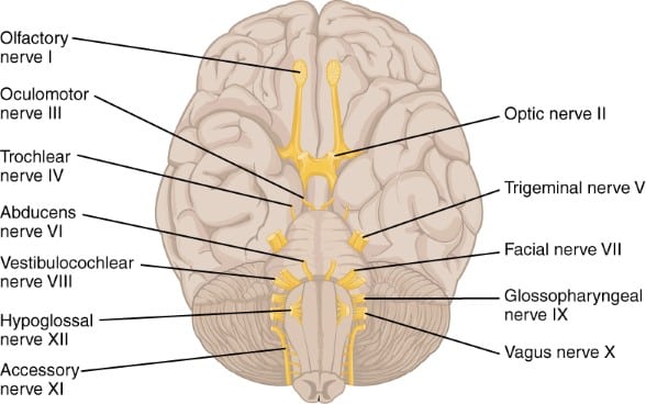
Cranial Nerves (source Anatomy & Physiology OpenStax College; Download for free at http://cnx.org/content/col11496/latest/)

Study Tips Neuroanatomy are concrete additions to your study strategy, First of all to make your study more successful and at the same time more fun with optimal use of the resources

Study Tip  Neuroanatomy 1.

First of all: Visualize neuroanatomy. Teaching assistants(=mentors) have passed the first course successfully and have spent a lot of time visualizing the neuroanatomy.

Visualize in a way that you enjoy and ‘ll simultaneously learn as you have fun making the visualization. You can have a look at the page Visualize your knowledge and let the work of members of our learning community inspire you.

Study Tip Neuroanatomy 2.

Furthermore you can use the resources in the Virtual Lab, Unit A: Neuroanatomy to get more information on core concepts of neuroanatomy.

Study Tip Neuroanatomy 3.

Let websites help you in visualizing neuroanatomy, Sylvius digital atlas is an especially relevant tool. But other websites can be very useful too. For example: for information on the optic tract   (5th image of the tracts presented) and location of the internal capsule and nuclei of deep gray matter in a human head.

Study Tip Neuroanatomy 4.

The textbook Neuroscience 2nd edition is on the NCBI bookshelf. It is a very useful resource because it has relevant images. For example  figure 1.121.13 and 1.14 for images on the lobes of the forebrain.

Study Tip Neuroanatomy 5.

There are general resources on the Virtual Lab, General Resources.

General Resources

and of course several Free online neuroscience textbooks. (Please be careful with what you share, avoid copyright infringement.)

Study Tip Neuroanatomy 6.

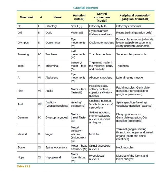
Here is a mnemonic to remember the Cranial Nerves: On Old Olympus Towering Tops A Fin And German Viewed Some Hops. You can find it in the Free, Open Source Table 13.3 from the OpenStax Book: Anatomy and Physiology

cranial nerves mnemonic
Cranial Nerves mnemonic (source Anatomy & Physiology OpenStax College; Download for free at http://cnx.org/content/col11496/latest/)


General Study Tips

General Study Tips


learn - pixabay cc0
Learn – Pixabay CC0

General Study Tips. Concrete additions to your study strategy to make your study more successful and more fun with optimal use of the resources.

General Study Tip 1.

I advise you to take several attempts at quizzes and exams before the deadline has passed, even if you haven’t watched all the videos. You have an unlimited number of quiz attempts (but may only retake a quiz 3 times in an 8-hour time frame).

The Quizzes are learning material. In  Coursera blog  the third tip for mastery learning is:

Practice, practice, practice! Take and re-take the quizzes available to you, look for extra-practice problems, create practice problems with your study group, etc. Think carefully through each attempt so that you’re getting the most out of your practice.

Use quizzes and exams for learning, not just for acquiring grades.

General Study Tip 2.

The learning goals summarize the learning material presented in the tutorial. Use them to learn for a quiz or an exam 😉

General Study Tip 3.

If you go to the tutorial videos you can find a list of downloads connected with that video at the right hand side of the screen just below the video. (See the instruction on how to find the tutorial notes and transcripts on the Medical Neuroscience Bulletin Board.) Here you can find a transcript of the video. You can download the transcript of the video in Notebook and use the material in your study, preparing for quizzes, while taking quizzes an while evaluating your quiz score. You can easily merge all transcripts in one document and make an Pdf for yourself that is easy to search.

General Study Tip 4.

A very useful tip by TA Nicole Foster on taking notes:

Take notes
Take Notes

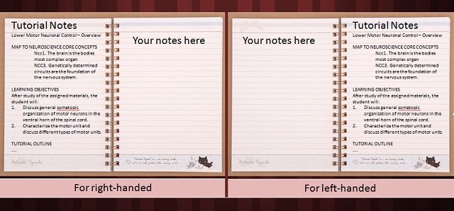
What I did when I took the course was print the tutorial notes. Dr. White mostly goes in the same order in the video. Give them a quick going over so you are familiar with the organization and content. Punch holes in the opposite side of what you normally would (if and only if you are right handed; if left. . . . punch holes as normal). Stick them into a binder backwards. That way, when you are on page 1, you have the back of page 2 to write on with your right hand. Take notes as you watch the videos. Don’t waste your time on things you already know.

If you have questions on a specific video, pause the video and write it down in a different color or with some symbol. Watch the rest of the videos. If the question is answered later on, go back and write that answer in. If not, do a book or internet search to see if you can track down the answer. If not, find a thread in the course forums that deals with that topic . . . . or start your own thread.

 General Study Tip 5.

Doing quizzes in the past I found that I missed key terms during the lectures. I did not understand questions and had to look things up in the textbook. If you do not have the textbook, this might help:

General Study Tip 6.

Most of the figures of the textbook are protected by copyright and only available in the textbook itself. Some are to be found in the second edition of the textbook on the NCBI  bookshelf.  They can be viewed but not shared.

You can make copies from images shown in the tutorials (for personal use only) by using “ctrl+alt+print screen,” then editing the images in a photo editor.

General Study Tip 7.

In the Virtual Lab: General Resources  you can find the link to the site: 3-D Brain App

This an extremely useful site, if you go to the 3-D brain you can look at separate brain structures. Not only that, case studies, associated functions, associated cognitive disorders, and associated with damage give a wealth of useful information. Make good use of this.

General Study Tip 8.

When reviewing the material of the course the Summary of Pathways  (2020, Neuroscience 5th and 6th ed.) This is a summary for the Final Exam of the course. Images in the Summary come from Anatomy & Physiology (OpenStax College .© 1999-2016, Rice University) is very useful.. You can use this material for educational purposes but remember to give the source. You can look at Virtual Lab, General Resources to find out how to do that.