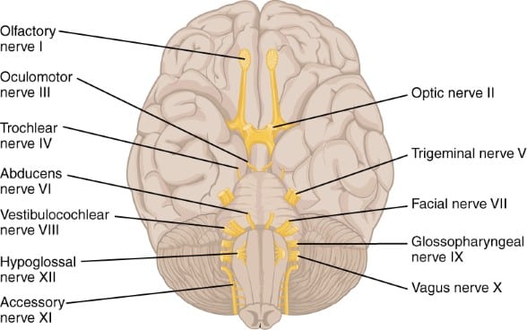
Cranial Nerves (source Anatomy & Physiology OpenStax College; Download for free at http://cnx.org/content/col11496/latest/)
Study Tips Neuroanatomy are concrete additions to your study strategy, First of all to make your study more successful and at the same time more fun with optimal use of the resources
Study Tip Neuroanatomy 1.
First of all: Visualize neuroanatomy. Teaching assistants(=mentors) have passed the first course successfully and have spent a lot of time visualizing the neuroanatomy.
Visualize in a way that you enjoy and ‘ll simultaneously learn as you have fun making the visualization. You can have a look at the page Visualize your knowledge and let the work of members of our learning community inspire you.
Study Tip Neuroanatomy 2.
Furthermore you can use the resources in the Virtual Lab, Unit A: Neuroanatomy to get more information on core concepts of neuroanatomy.
Study Tip Neuroanatomy 3.
Let websites help you in visualizing neuroanatomy, Sylvius digital atlas is an especially relevant tool. But other websites can be very useful too. For example: for information on the optic tract (5th image of the tracts presented) and location of the internal capsule and nuclei of deep gray matter in a human head.
Study Tip Neuroanatomy 4.
The textbook Neuroscience 2nd edition is on the NCBI bookshelf. It is a very useful resource because it has relevant images. For example figure 1.12, 1.13 and 1.14 for images on the lobes of the forebrain.
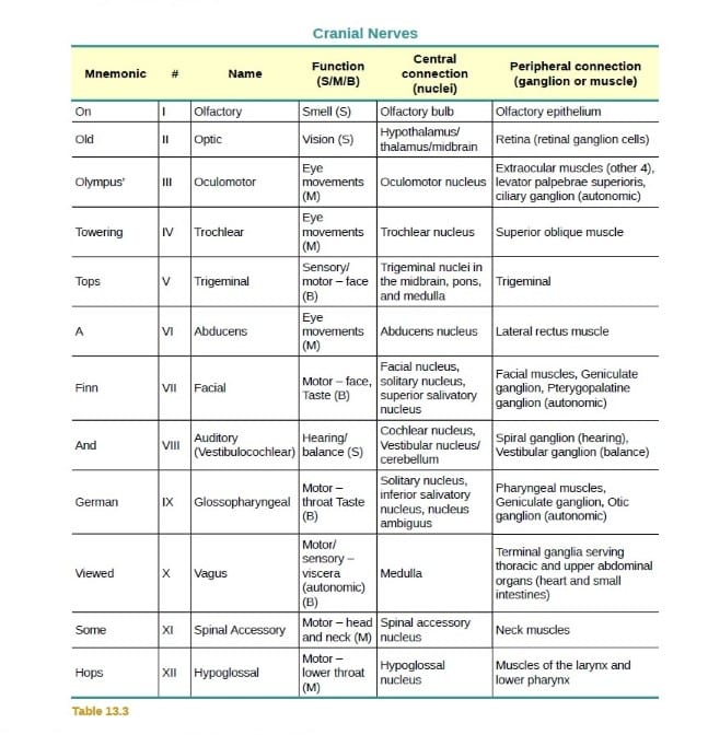
Here is a mnemonic to remember the Cranial Nerves: On Old Olympus Towering Tops A Fin And German Viewed Some Hops. You can find it in the Free, Open Source Table 13.3 from the OpenStax Book: Anatomy and Physiology
Cranial Nerves mnemonic (source Anatomy & Physiology OpenStax College; Download for free at http://cnx.org/content/col11496/latest/)
“Visualize your knowledge”- prof. Len White, Tutorial Video Your Part! at 07:15
The Course Medical Neuroscience
Welcome to the Learn Medical Neuroscience website. Most of all this neuroscience website supports members of the learning community of the Coursera course “Medical Neuroscience“. Prof. Len White from Duke University teaches this course. Other Neuroscience learners are welcome of course but they are not primary target group of the website.
The website Learn Medical Neuroscience
The information presented here is intended to strengthen a learner’s knowledge and understanding of course content as well as to provide information about useful learning strategies. As such, all of the information herein is for supplementary strengthening purposes and is not intended to exceed the scope of the course.
This website was made and is maintained by Ellen Vos-Wisse, an active member ( Mentor) of the Medical Neuroscience learning community. Find out more about Ellen on the contact page of this website where she tells you more about her background and her connection to Medical Neuroscience.
Structure of the website Learn Medical Neuroscience
This is the place for more information on the Global Medical Neuroscience class. Here you can find information on how to use the discussion forum on the course website, examples from pictures of #GetNeuro challenges from the course that are shared on Instagram and the link to a highly active Facebook group for the course. There are nice pictures on ‘Medical Neuroscience behind the scenes’. The News page presents news about the course and general neuroscience news, all relevant to the learning community.
Actually this is the core of the website. You can find a list of links to relevant websites on the internet all about the brain in the Virtual Lab . The Virtual Lab has 7 sub pages. The structure of the Medical Neuroscience course is the foundation of the Virtual Lab. The page Virtual Lab explains the structure of the content of this part of the website. The featured videos are a nice part of the Virtual Lab. Featured videos are short videos on the field of Neuroscience relevant to the information of the content of the page. They are not always directly linked to the content of the sub page of the virtual lab.They form a complement to the core material of the course and give additional insight.
At this page you can find more links to more websites. The sites that this part of the website links to do not explain course content. This page presents links to websites you can use to review your Neuroscience knowledge. This page also contains Neuroscience book recommendations. In addition to that there is a link to the Summary of Pathways Medical Neuroscience. This Summary of Pathways is a document, free of copyright, made especially for this course. You can download it directly from the site.
The Neurological Exam is a part of the website with links to sites and videos explaining the neurological exam. There is a link to the especially relevant site NeuroLogical Cases of the University of Utah. This is very systematic and educationally thorough site to learn the Neurological Exam. Other resources are also present on this page. Such as ‘Clinical Neurology Videos’ a wonderful resource, specifically created to enhance and facilitate the study of Neurology. You can also have a look at the cases in the paragraph: ‘Dealing with Neurological Constraints’. There you find inspirational material that shows that people can adapt to great neurological constraints and live a full life despite these constraints.
At the right side of the screen (not on pages of the Virtual Lab and the page Neurological Exam) you see News and under that pages with Study Strategy and Study Tips. These pages have proven very useful to learners in the past. You can check them out and use them to your advantage. Last but certainly not least there is the Course Bulletin Board. There you can find information that is important when you take the course like: Where are the Tutorial Notes? What happens to my grades when I switch to a next session?
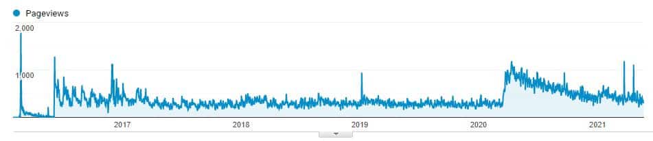
The website has been well used since the start the site in 2016. On May 23 2021 there have been 160,495 users of the site and 696,501 pageviews. You can see in the graph below that the use of the site intensified during the pandemic.
Number of pageviews in time of the website www.learnmedicalneuroscience.nl
Featured video of the page Neuroanatomy: “Visualizing the brain as a universe of synapses” by Stanford Medicine. For more information see the description of the method.
Neuroanatomy in the course Medical Neuroscience
On this page you can find the links to resources on Neuroanatomy. These are not only relevant in your study of the unit Neuroanatomy in the course Medical Neuroscience but are also very useful in later units of the course, Neuroanatomy is really the foundation in the course. It is a good idea to use the resources throughout the course.
3-D Brain App, Genes to Cognition Online by Cold Spring Harbor Laboratory. A very useful 3D model of the brain. It has descriptions of various brain structures, associated functions, associated cognitive disorders, research reviews etc. It is useful for the preparation of Quizzes and especially relevant for the Comprehensive Final Exam. It was accessible with the internet browser, but that site used Flash, it does not work anymore. You have to download the App. The The You can get the free 3D Brain app from Google play and Windows Phone and the App Store from Apple.
The Human Brain by Anatomie-Amsterdam : Brain atlas for educational purposes, with clear interactive images. Includes external views of the brain and cerebellum, cerebrum slices , white matter, tracts and MRI scans. (Move the cursor over the image to see the labels).
3D Brain by BrainFacts.org. Interactive brain model with clear and helpful structure descriptions.
MRI Atlas of the Brain. An interactive presentation of the anatomical structures, by Javad Hekmat-panah, MD, Professor of Neurosurgery, Neurology and Cancer Research University of Chicago.
Allen Brain Atlas: Contains several free online atlases of the human and mouse brain adult and developing brains, published by the Allen Institute (Paul Allen is one of the founders of Microsoft). The data are heavily geared towards gene expression, but the reference atlases are quite usable and useful. Have a look at the Allan Brain Atlas tutorials.
Anatomy of the Brain by University of British Columbia with a Creative Commons License: web atlas, imaging, 3D reconstructions, stroke model, brain & behaviour, neurotutorials , movies.
The Unfixed Brain: YouTube video by University of Utah Brain Institute.
ZygoteBody Anatomy: create a free account and use the 3D Brain Model to learn
Brainbows — Mixing Colors to Map the Brain In this video from Vania Cao shows how scientists developed the “brainbow” technique to differentiate neurons by color, and it took third place in the 2014 Brain Awareness Video Contest of the SfN.
Non-Neural Cells of the Central Nervous System (CNS)
Neuroglia and the Brain. Video about glia, thelesser-known brain cells support and protect neurons. Brain Awareness Video Contest 2013 of the SfN. Video by secondary school students Yash Patel, Michelle Goffreda, Robby Vasen, and Kat Lin of High Technology High School, Lincroft, NJ.
Cranial Nerves Basics. A resource with mnemonics found by a former student (Mauro Mello Jr) in the course and which has been very useful to him, who says “I am doing a formal specialization in neuroscience and am still amazed with what I learned with Professor White… Without a shadow of doubt MNS has been the best course on neuroscience I have ever taken.” The video is a bit long but the bits that are interesting start at 12:02. Feel free to view the whole video.
Cranial nerves video from the Brain Awareness Video Contest 2011 of the SfN by Scarth Locke, a Samuel Merritt University graduate student, , along with associate professor Barb Puder.
The Blood Supply of the Brain and Spinal Cord. Go to Neuroscience 2nd edition at the NCBI bookshelf. Enter “The Blood Supply of the Brain and Spinal Cord” in the search box for this book. Figures 1.19, 1.20 and 1.21 are extremely useful.
Be careful do not violate copyright. You can share images from Anatomy & Physiology if you cite the source, including the download link (see Virtual Lab, General Resources). Featured videos: only YouTube and Vimeo videos with a “Share” button have been embedded. If the owner enables the Share button, it means that they allow others to embed the video. If the copyright owner of the video has made a mistake and does not want the video embedded despite the Share button, please inform me. I will remove the video immediately. -Ellen-
 A very useful 3D model of the brain. It has descriptions of various brain structures, associated functions, associated cognitive disorders, research reviews etc. It is useful for the preparation of Quizzes and especially relevant for the Comprehensive Final Exam. It was accessible with the internet browser, but that site used Flash, it does not work anymore. You have to download the App. The The You can get the free 3D Brain app from Google play and Windows Phone and the App Store from Apple.